










Surgical Treatment at Swift Access Health

Melanoma

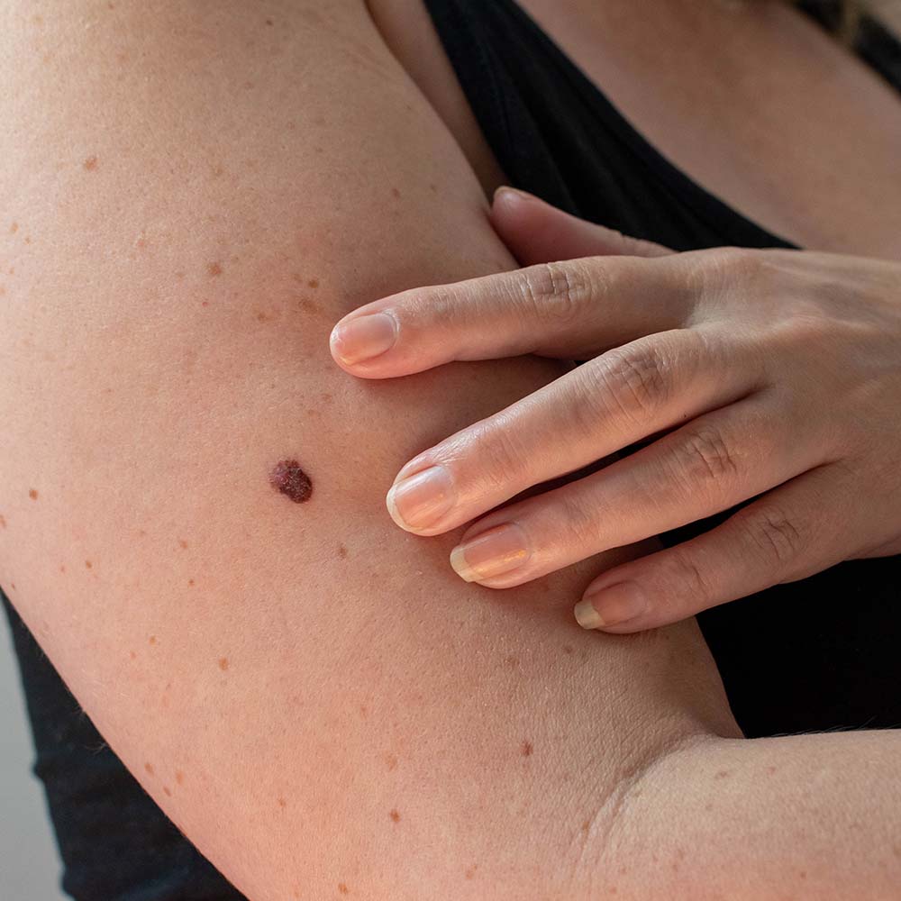
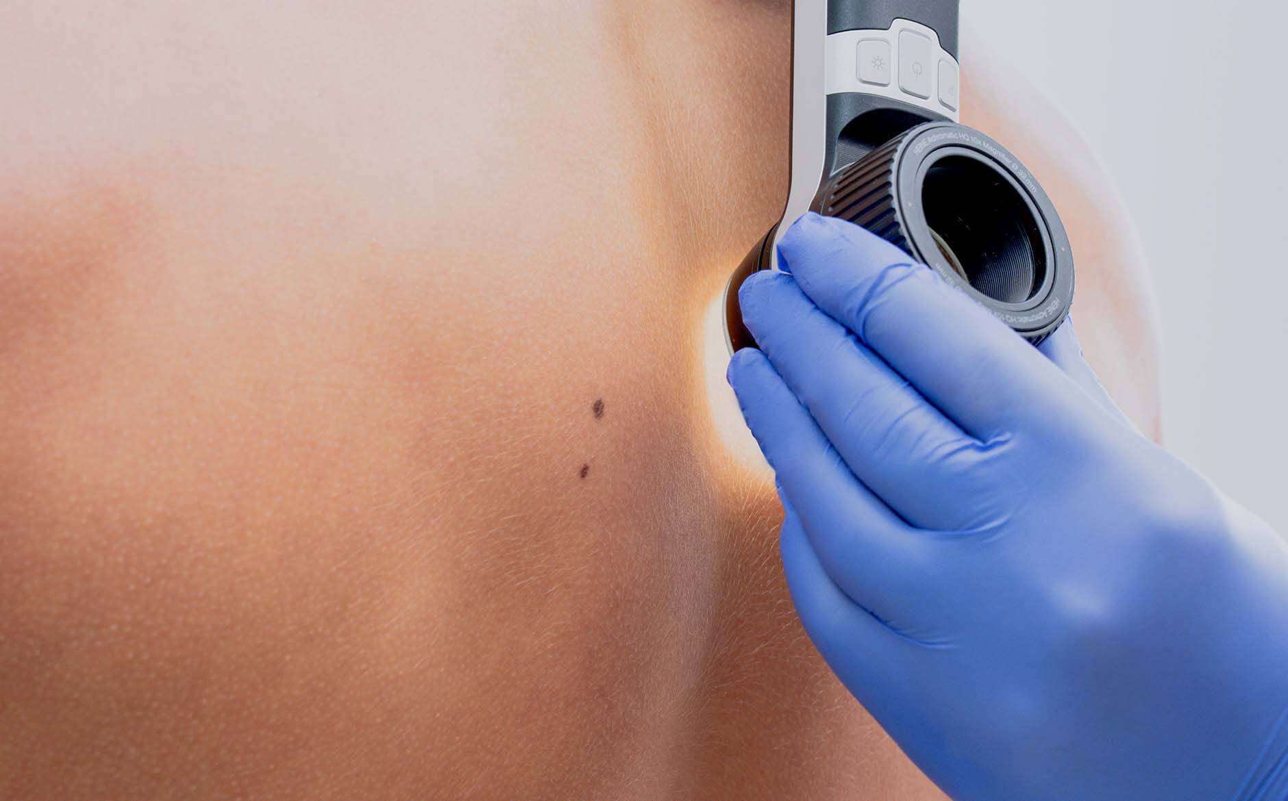
Melanoma is a form of skin cancer that develops from the melanocytes — the cells responsible for producing pigment. It is less common than basal cell or squamous cell carcinoma, but significantly more serious due to its capacity to spread to lymph nodes and distant organs if not detected and treated early. Melanoma can arise in an existing mole or appear as a new lesion, often with features of asymmetry, irregular border, multiple colours, or change over time.

At Swift Access Health, melanoma is managed by consultant dermatologists and plastic surgeons who are experts in skin cancer and are members of both NHS and private skin cancer multi-disciplinary teams. Management involves wide local excision with appropriate margins and, in suitable cases, sentinel lymph node biopsy to determine whether the cancer has spread. Advanced disease is managed in collaboration with oncology colleagues for immunotherapy or targeted therapy where required.

Book Consultation

Info

Time

30–60 minutes for wide local excision; sentinel lymph node biopsy adds time

Results

Complete surgical removal of early melanoma is curative in the majority of cases

Risks

Bleeding, infection, scarring, lymphoedema; recurrence risk dependent on stage — discussed fully

Recovery

7–14 days for initial wound healing; further treatment if sentinel node is positive

Anaesthetic

Local anaesthetic for most cases; general anaesthetic occasionally used for complex sites

Aftercare

Wound care, sun avoidance, regular skin checks, and structured oncological follow-up

Why Choose Swift Access Health

Benefits of Specialist Melanoma Surgery

Surgery by a consultant dermatologist or plastic surgeon with specialist expertise in melanoma ensures accurate staging, technically precise excision, and coordinated care across the full treatment pathway.

  • Wide local excision in line with NICE-guideline margins to minimise local recurrence
  • Sentinel lymph node biopsy performed where indicated to accurately stage the disease
  • Reconstructive expertise to restore function and appearance following wide excision
  • Multidisciplinary team approach with oncology for advanced disease requiring systemic treatment
  • Structured long-term surveillance with regular skin checks and clinical review
Book Consultation
Your Patient Journey

1. Your
Consultation

Your surgeon will review any existing biopsy report, examine the primary site and regional lymph nodes, and arrange any further imaging or staging investigations required. The surgical plan — including excision margins, closure technique or reconstruction, and the role of sentinel lymph node biopsy — will be explained clearly. A personalised treatment plan is agreed before consent is obtained.


Your Patient Journey

2. Your
Treatment

The melanoma is excised with an appropriate margin of healthy skin determined by the tumour’s Breslow thickness. Where sentinel lymph node biopsy is indicated, the draining lymph node is identified and sampled to check for microscopic spread. The wound is closed directly or reconstructed using a skin flap or graft as required. All tissue is sent for histological examination.

Your Patient Journey

3. Aftercare

Following surgery, the wound is kept clean and dry and sutures are removed after 7–14 days. Histology results are reviewed at a follow-up appointment and the next steps — whether surveillance or referral for systemic treatment — are discussed. Long-term follow-up includes regular skin checks and, for higher-stage disease, imaging surveillance. Sun protection and skin self-examination are essential.

Frequently Asked Questions

  • Melanoma arises from melanocytes and is biologically more aggressive than basal cell or squamous cell carcinoma, with a greater capacity to spread through the lymphatic system and bloodstream to distant organs. Early detection and treatment are critical — survival rates for early-stage melanoma are excellent, but decline significantly once the disease has spread.

  • Use the ABCDE criteria: Asymmetry, irregular Border, multiple Colours (brown, black, red, white, or blue), Diameter greater than 6mm, and any Evolving change in size, shape, colour, or new symptoms such as bleeding or itching. Any changing or new lesion should be assessed by a dermatologist promptly.

  • Anyone with a lesion that has changed rapidly, that bleeds without trauma, or that a clinician has already identified as suspicious should be seen urgently. Patients with a confirmed melanoma diagnosis awaiting surgery should be prioritised for early operative planning.

  • Sentinel lymph node biopsy (SLNB) is a minimally invasive procedure that identifies and samples the first lymph node draining the melanoma site. If melanoma cells are present in the sentinel node, it indicates potential spread and informs decisions about further treatment. SLNB is recommended for melanomas of Breslow thickness 1mm or greater.

  • If sentinel lymph node biopsy confirms lymph node involvement, or imaging identifies distant spread, treatment moves from surgery alone to a combination of surgery and systemic therapy. Modern immunotherapy and targeted therapy drugs have significantly improved outcomes for patients with advanced melanoma.

  • Regular follow-up is essential following melanoma treatment. Frequency and duration depend on the stage of the disease. Early-stage melanomas are typically followed for 5 years with annual skin checks. Higher-stage disease requires more intensive surveillance. Patients are taught self-examination techniques and advised on sun protection measures.

Related Concerns

Concerns

Moles

Concerns

Skin Cancer