











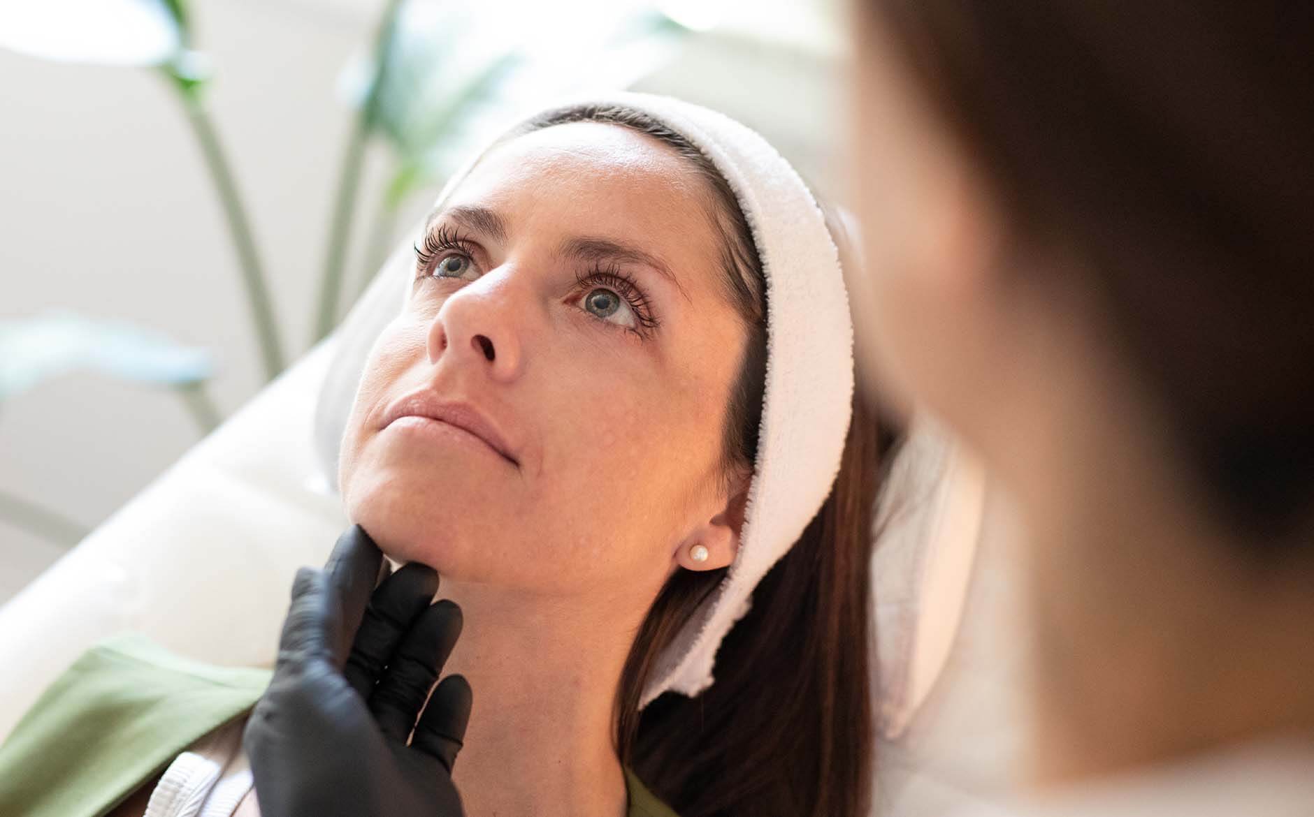

Diagnostic Procedure at Swift Access Health

Skin Biopsy

A skin biopsy is a minor surgical procedure in which a small sample of skin is removed and sent to a specialist pathology laboratory for microscopic analysis. It is the gold standard method for confirming the diagnosis of many skin conditions — from inflammatory rashes and lesions of uncertain cause to suspected skin cancers. The results guide treatment decisions and provide definitive diagnostic certainty that clinical examination alone cannot always offer.

Skin biopsies are performed by consultant dermatologists and plastic surgeons using the latest biopsy techniques relevant for each lesion type. The procedure is quick, performed under local anaesthetic, and causes minimal discomfort. Results are typically available within 7–14 days, at which point a follow-up appointment is arranged to discuss findings and agree next steps.

Book Consultation

Info

Time

15–30 minutes including anaesthetic and wound closure

Results

Definitive histological diagnosis within 7–14 working days

Risks

Minor bleeding, infection, scarring at biopsy site; rare allergic reaction to local anaesthetic

Recovery

Wound heals within 1–2 weeks; sutures removed after 7–14 days if required

Anaesthetic

Local anaesthetic injection — the procedure itself is painless

Aftercare

Keep wound clean and dry; avoid strenuous activity; attend follow-up for results and suture removal

Why Choose Swift Access Health

Benefits of Skin Biopsy

A skin biopsy performed by a specialist and analysed by an expert dermatopathologist provides definitive diagnostic answers — enabling the right treatment to start promptly and with confidence.

  • Performed by an expert consultant dermatologist or plastic surgeon
  • Choice of biopsy technique matched to the lesion type — punch, shave, incisional, or excisional
  • Specimens analysed by experienced dermatopathologists with specialist expertise in skin disease
  • Fast turnaround with results typically available within 7–14 working days
  • Follow-up appointment included to discuss results and agree on next steps
  • Urgent processing available for suspected skin cancers where clinical urgency dictates
Book Consultation
Your Patient Journey

1. Your
Consultation

Your clinician will explain why a biopsy is being recommended, what type of biopsy will be performed, and what the results may indicate. The risks of the procedure are discussed and consent is obtained. The target lesion is examined and the procedure is planned, including the choice of technique and the expected scar.


Your Patient Journey

2. Your
Treatment

Local anaesthetic is injected around the lesion to numb the area completely. The biopsy is then taken using the appropriate technique — most commonly a punch biopsy (using a circular cutting tool to remove a core of skin), shave biopsy (removing a surface layer of skin), or excisional biopsy (removing the lesion in its entirety). A small dressing is applied and the sample is sent to the laboratory.

Your Patient Journey

3. Aftercare

The wound should be kept clean and dry for 48 hours. After this, it can be gently cleansed in the shower. Sutures are removed after 7–14 days if placed. A follow-up appointment is arranged to discuss the histology results. Treatment recommendations are made based on the findings — which may include further surgery, medical treatment, or reassurance and surveillance.

Frequently Asked Questions

  • The local anaesthetic injection causes a brief stinging sensation, after which the biopsy site is completely numb and the procedure is painless. Some mild tenderness around the biopsy site is normal in the days following, which resolves quickly and is managed with standard over-the-counter pain relief.

  • A punch biopsy removes a small cylindrical core of full-thickness skin using a circular cutting tool — ideal for inflammatory conditions and pigmented lesions. A shave biopsy removes a superficial sample using a blade — suitable for raised, benign-appearing lesions. An incisional biopsy samples a portion of a larger lesion. An excisional biopsy removes the entire lesion with a margin — combining diagnosis and treatment.

  • Results are typically available within 7–14 working days from the date of the biopsy. If the laboratory requires additional specialist stains or second-opinion reporting, this may extend the timeline slightly. Your clinician will inform you if any delay is expected. Results are never communicated by letter alone — a follow-up appointment is always arranged.

  • All skin biopsies leave a small scar. The size depends on the biopsy technique and the size of the sample taken. Punch and shave biopsies typically produce very small, inconspicuous marks. Excisional biopsies leave a linear scar that fades over 12 months. Your clinician will place the biopsy site to minimise scar visibility wherever possible.

  • Sampling error — where the biopsy does not capture representative tissue — is occasionally possible, particularly with large or heterogeneous lesions. Your consultant dermatologist selects the most informative site for biopsy to minimise this risk. If the histology result does not correlate with the clinical picture, repeat biopsy or further investigation may be recommended.

  • Skin biopsy is used to diagnose inflammatory conditions such as psoriasis, lichen planus, lupus, and dermatitis; bullous (blistering) diseases; pigmented lesions including atypical naevi and melanoma; suspected skin cancers including BCC, SCC, and lymphoma; and unusual or persistent rashes that have not responded to empirical treatment.

Related Treatments

Treatments

Allergy Testing

Treatments

Lipoma Removal