











Diagnostic Service at Swift Access Health

Mole Mapping

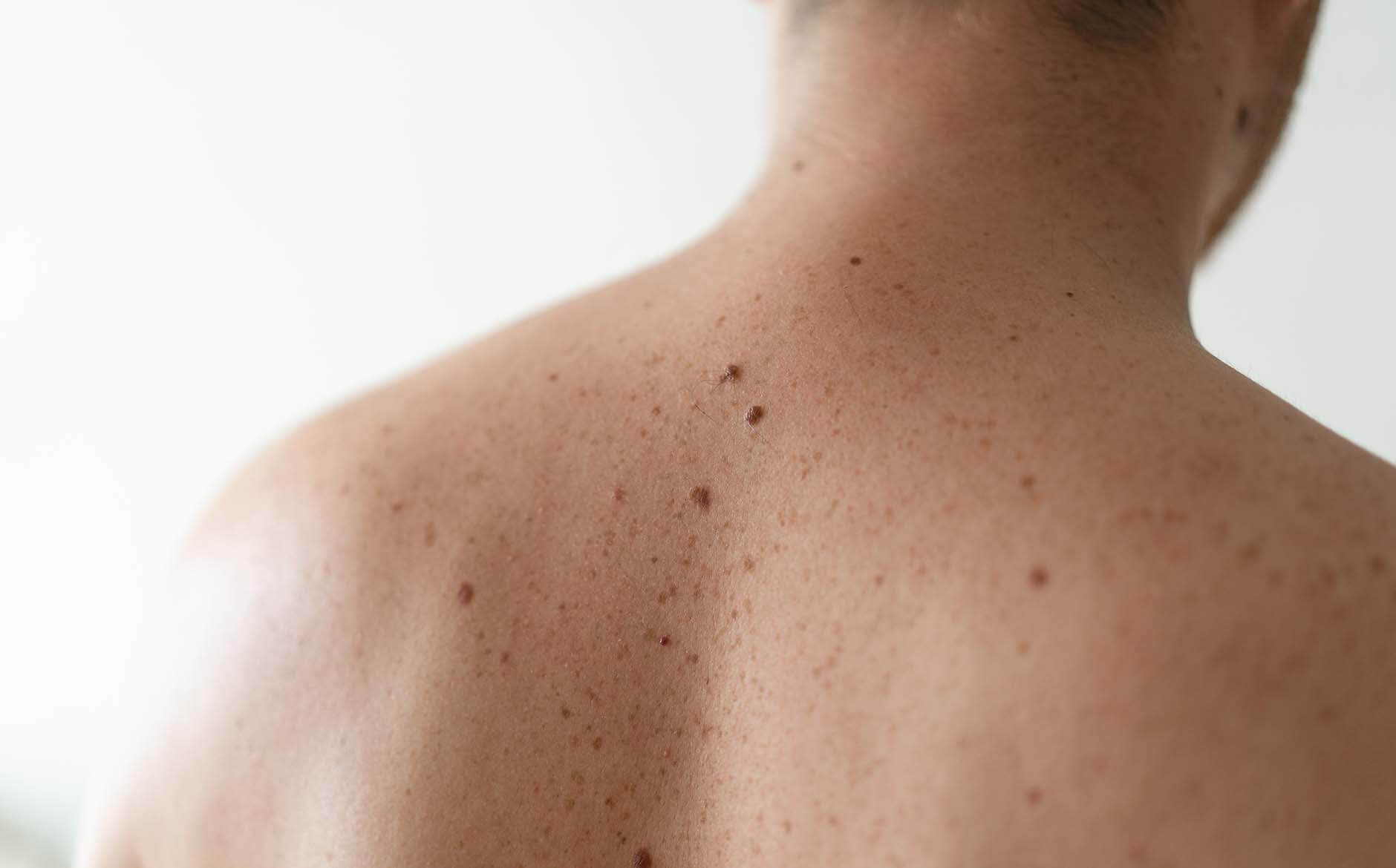
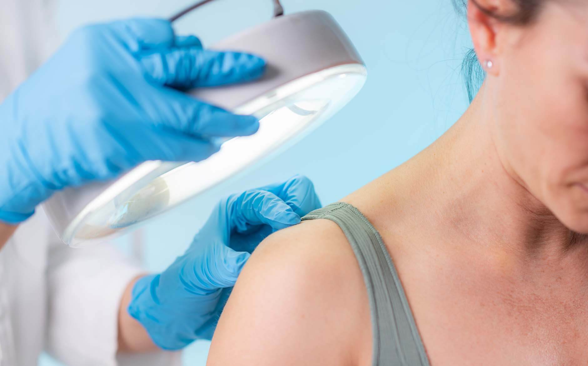
Mole mapping is a specialist surveillance service for patients at increased risk of melanoma or those with multiple moles, atypical naevi, or a personal or family history of skin cancer. The service involves a systematic clinical examination by a consultant dermatologist, combined with dermoscopic assessment and standardised clinical photography of individual lesions so they can be tracked over time. Images are recorded and stored, enabling precise comparison at future appointments to detect any change in size, shape, structure, or colour over time.

At Swift Access Health, mole mapping is performed and interpreted by a consultant dermatologist using high-resolution dermoscopy and clinical photography. This is a consultant-led service — not an automated or technology-only assessment, and provides a diagnosis and clear plan on the day, unlike most other clinics.

Photographs are reviewed in the context of a full clinical examination, and any lesion showing worrying features is assessed and managed promptly. The goal is early detection of abnormal moles or melanoma at the most treatable stage.

Book Consultation

Info

Time

30-45 minutes for full-body assessment; 30 minutes for follow-up reviews

Results

Comprehensive baseline record of all moles; early detection of any concerning changes at review

Risks

None from the mapping procedure itself; any suspicious lesion identified is biopsied or excised

Recovery

No recovery required — non-invasive photographic assessment

Anaesthetic

None required for the mapping procedure

Aftercare

Structured review intervals agreed; sun protection and self-examination guidance provided

Why Choose Swift Access Health

Benefits of Consultant-Led Mole Mapping

Consultant dermatologist-led mole mapping provides the most thorough and clinically meaningful surveillance for patients at increased melanoma risk — combining human expertise with systematic photographic monitoring.

  • Performed and interpreted by a consultant dermatologist which remains the gold standard — not a technician or AI-only system
  • Same-day diagnosis – not waiting for days or weeks like other clinics
  • High-resolution dermoscopic photography of individual lesions for precise lesion-level comparison
  • Full clinical examination at every appointment — not a photography-only service
  • Any suspicious lesion identified at mapping can be assessed and acted upon in the same appointment
  • Structured review intervals agreed based on individual risk profile — typically 6 or 12 months
  • Clear sun protection, self-examination, and lifestyle guidance to support long-term skin health
Book Consultation
Your Patient Journey

1. Your
Consultation

Your initial mole mapping appointment begins with a clinical consultation in which your dermatologist takes a full history — including personal and family history of skin cancer, skin type, sun exposure, and any moles that have recently changed. A total body skin examination is performed, with dermoscopic assessment of all lesions above a size or complexity threshold. Photographs are taken and stored.


Your Patient Journey

2. Your
Treatment

This is a diagnostic and surveillance service — not a treatment procedure. However, any lesion that is considered suspicious on clinical or dermoscopic assessment will be biopsied or excised at an expedited follow-up appointment. Your dermatologist will explain any such recommendation clearly, with a rationale based on the specific features of concern.

Your Patient Journey

3. Aftercare

Following your mole mapping appointment, you will receive guidance on skin self-examination — what to look for between appointments and when to seek an earlier review. A structured follow-up interval is agreed, typically every 6–12 months depending on your risk profile. Sun protection with SPF 50+ and protective clothing is strongly recommended as part of your long-term skin health strategy.

Frequently Asked Questions

  • Mole mapping is recommended for all patients wanting to be proactive with their skin health. In addition, it is invaluable for those with more than 50 moles, a history of atypical or dysplastic naevi, a personal or family history of melanoma, fair skin with significant cumulative sun exposure, or any moles that have previously required removal or close monitoring. Your dermatologist can advise on whether the service is appropriate for your risk profile.

  • A routine skin check involves clinical examination and dermoscopy at a single point in time. Mole mapping adds the dimension of serial photography, enabling precise comparison of individual lesions over time. Change — even subtle change — that would be impossible to detect by clinical examination alone becomes visible when images from successive appointments are compared side by side.

  • Review intervals depend on individual risk. Most patients are reviewed annually. Those with higher-risk profiles, multiple atypical naevi, or a history of melanoma may benefit from 6-monthly reviews. Your dermatologist will agree the most appropriate frequency for you at your initial assessment.

  • Any lesion identified as suspicious at mole mapping can be biopsied or excised promptly. All excised specimens are sent for histological analysis. Patients are never left waiting with an unresolved concern.

  • Attend with clean skin and no make-up, fake tan, or nail polish on the day of your appointment. Wear or bring loose clothing that allows easy access to all skin areas. Bring any previous mole mapping records or skin examination reports from other providers so that your dermatologist can review these alongside the new images.

Related Treatments

Treatments

Mole Checks

Treatments

Mole Removal